Precut, prestained tissue segments may be advantageous in keratoconus surgery

Article originally published on Healio, Nov. 16, 2021.


Figure 1a. Precut and prestained CAIRS immediately after preparation in an eye bank setting. Figure 1b. The donor material is shipped in storage solution, rinsed with saline and then implanted.

Corneal allogenic intrastromal ring segments have been described by Jacob and colleagues for the treatment of keratoconus.

They consist of semicircular pieces of donor corneal stroma that may be surgically implanted to flatten, stiffen and stabilize the shape of the recipient cornea.

Early corneal allogenic intrastromal ring segments (CAIRS) protocols used donor segments prepared immediately before implantation, at the time of the surgery. However, because this process involves multiple steps, it may add significantly to the overall procedure time. In addition, any problems encountered during segment creation could jeopardize the ability to complete the operation. Therefore, it may be advantageous to prepare the donor material in advance.

Similar considerations have inspired the recent utilization of so-called “patient-ready” tissue for Descemet’s membrane endothelial keratoplasty, ie, donor endothelium that has been prestripped, prestained and preloaded. Since this concept was introduced in 2016, studies have demonstrated robust stain retention and tissue viability, with surgical outcomes comparable to those in which the tissue is prepared intraoperatively. Patient-ready tissue may also facilitate the uptake of DMEK by novel surgeons by diminishing the anxiety and risks associated with graft preparation.

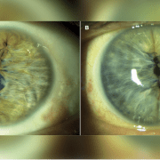
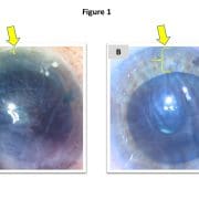
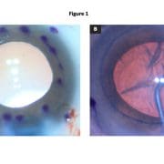
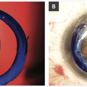
To date, we have performed CAIRS in 16 eyes of 15 patients with keratoconus using precut, prestained tissue. These patient-ready segments are prepared within an eye bank setting 48 to 72 hours before surgery. A donor corneoscleral button is debrided of epithelium and endothelium using a surgical sponge and then mounted on a Teflon cutting block. A dedicated CAIRS trephine (Jacob CAIRS trephine, Madhu Instruments) is used to create a circular tissue ring that is bisected via surgical knife into two semicircular segments. The option also exists to supply it as a full ring to allow customization of arc length by the surgeon. Both segments are submerged in 0.06% trypan blue (VisionBlue, DORC International) for 3 minutes until a dark blue tissue stain is obtained and then replaced in storage solution and photographed (Figure 1a). At the time of surgery, the segments are removed from storage, irrigated with saline, dehydrated atop a surgical sponge and implanted (Figure 1b).

Since transitioning to patient-ready CAIRS, no additional intraoperative or postoperative complications have been experienced. As with prestripped and prestained DMEK grafts, the tissue retains its blue stain throughout the duration of the procedure but fades completely by several days postoperatively.

Advantages of patient-ready CAIRS may include reduced surgical time, flattened surgical learning curve and increased consistency of product. As a result, this may become a preferred option for tissue preparation, especially for surgeons starting with the technique.

References:

  • Jacob S, et al. J Refract Surg. 2018;doi:10.3928/1081597X-20180223-01.
  • Parekh M, et al. Am J Ophthalmol. 2016;doi:10.1016/j.ajo.2016.03.048.
  • Parker JS, et al. J Cataract Refract Surg. 2021;doi:10.1097/j.jcrs.0000000000000316.
  • Parker JS, et al. J Cataract Refract Surg. 2021;doi:10.1097/j.jcrs.0000000000000582.
  • Potts LB, et al. Cornea. 2020;doi:10.1097/ICO.0000000000002400.
  • Zafar S, et al. Clin Ophthalmol. 2019;doi:10.2147/OPTH.S212871.

For more information:

Hudson Tate, David Blackburn and Jack S. Parker, MD, PhD, can be reached at Parker Cornea, 700 18th St. South, Suite 503, Birmingham, AL 35233; emails: hdtate@crimson.ua.edu; davidblackb60@gmail.com; jack.parker@gmail.com.

Continue reading on Healio.


Short learning curve results in success with pinhole pupilloplasty

Article originally published on Healio, July 9, 2021.

Pinhole pupilloplasty has helped change many lives. The idea of small-aperture optics is not only simple but advantageous in cases of high astigmatism.

Jack S. Parker, MD, PhD, of Parker Cornea and the Netherlands Institute for Innovative Ocular Surgery (NIIOS), is my guest columnist this month, and he will explain with his co-author how to master pinhole pupilloplasty (PPP).

The terminology of pinhole pupilloplasty was suggested to me by David Chang, and I will always be grateful to him for coming up with such a suitable name. PPP can be done with any technique of iris suturing. Since McCannel started iris suturing and then Siepser took it to another level, so many others have pioneered in this area. Ogawa brought in the cerclage, and Ike Ahmed brought in the McAhmed suturing technique. Priya Narang helped pioneer single-pass four-throw pupilloplasty, for which I am indebted. The four-throw technique basically got us out of the locking system that was being used in all the iris suturing techniques. This made the whole surgery of iris suturing easy and possible by any surgeon. When we perform PPP, we generally use the single-pass four-throw pupilloplasty technique, although one can use any technique of their choice.

Pinhole pupilloplasty (PPP) was recently described for the treatment of severe corneal astigmatism. Pinhole apertures block stray distorted light rays and confer enhanced depth of focus. For this reason, pinhole IOLs are readily available in many places globally but not in the United States, where the FDA has yet to approve them. Therefore, Dr. Agarwal published a technique for recreating the pinhole aperture naturally by suturing the iris to produce a pinhole-sized pupil.

Initial publications with this technique are impressive: Eyes with more than 10 D and even more than 20 D of irregular (often fluctuating or unstable) astigmatism from prior penetrating keratoplasty or radial keratotomy are improved, in many cases achieving better than 20/40 (0.5) uncorrected vision. Further, PPP is cost-effective, uses already widely available and FDA-approved materials (suture), and is easily reversible without return to surgery, for example, by lasering the iris stitches, which can be done in clinic.

These advantages inspired us to start with PPP, and here, we describe some difficulties and insights acquired during our learning curve with the procedure.

1. Size is important. Ideal for most patients is 1.5 mm. Any larger, and the benefit of the pinhole aperture is reduced (Figure 1a). If smaller, visual acuity can become limited by diffraction (Figure 1b). Consider erring on the side of “too small” because the pupil may be subsequently enlarged via YAG laser postoperatively.

Pupil size
Figure 1. Appropriately sized 1.5-mm pupil of a patient with 20/20 uncorrected vision after PPP (a) compared with a less than 1-mm pupil of a patient with 20/80 vision after PPP (b), likely limited at least partially by diffraction through the aperture, which is too small.Source: Eric Kim, BS, BA, and Jack S. Parker, MD, PhD

2. Centration is important, too. The pinhole aperture should be fashioned around the first Purkinje image as a reference marker. A decentered pinhole-sized pupil is as optically offensive as a decentered multifocal IOL and reliably results in suboptimal visual outcomes (Figure 2).

pupil centering
Figure 2. Temporally decentered pupil (a) vs. properly centered PPP (b).

3. Use bracketing sutures. Both sides of the pupil must be sutured. Otherwise, when the preoperative or intraoperative miotic agent wears off, the patient will experience asymmetrical re-dilation (Figure 3).

sutured on only one side
Figure 3. Well-centered and well-sized PPP; however, notice that it is sutured on only one side (yellow arrow). Postoperatively, this patient experienced asymmetrical re-dilation, resulting in a tear drop-shaped, excessively large pupil. To prevent this, sutures should have been placed on either side of the pupil.

4. Suturing is facilitated by micro-grasping forceps. Dr. Agarwal originally described a “railroad technique” in which the suturing needle passes through the distal iris, a 26-gauge needle passes through the proximal iris, and the two are docked together and withdrawn from the eye. However, the 26-gauge needle often creates large needle track holes in the proximal iris, which are cosmetically and perhaps functionally detrimental (Figure 4a). So, we have replaced the 26-gauge needle with 25-gauge micro-grasping forceps, which may be used to hold and withdraw the suturing needle from the eye (Figure 4b). At present, Dr. Agarwal uses a 30-gauge needle, which is smaller and less traumatic to the iris.

Needle track holes
Figure 4. Needle track holes are visible in the iris surrounding the PPP (a), which are the product of using a 26-gauge needle to dock the suturing needle. When micro-grasping forceps are used instead, the cosmetic result is much improved (b).

5. Consider combining PPP with phacoemulsification and IOL implantation. Best for surgeons starting with PPP are phakic eyes also needing cataract surgery. In such cases, the phaco alone will likely render some benefit, obviating the need to execute the perfect PPP to satisfy patient expectations.

PPP has been a significant addition to our practice because, for the first time, we are able to offer some solution for patients with severe, irregular, fluctuating astigmatism (for example, from prior RK or PK) who are otherwise unable or unwilling to tolerate hard contact lenses. The procedure is quick and straightforward. It can be incorporated across most settings with ease as there is no need for specialized equipment.

Further, the operation is easy to recommend because, from their personal experience with eye exams, patients intuitively understand the visual improvement that they derive from pinholes. For doubters, a short course of topical pilocarpine may be tried preoperatively. Then, patients may be asked whether the resulting miosis improved their vision and, if so, whether they would like to have this effect recreated surgically.

PPP has predictable results that can be easily demonstrated preoperatively, allowing patients to have a tangible understanding and expectation of the procedure. It is also safe, reversible and highly effective for patients with few other options. After a short learning curve, it has become a regular surgical procedure at our practice, and most of our patients have reported great improvements without complications. As we continue to refine our technique, we expect even greater success with PPP.

Continue reading on Healio.


Parker cornea

VIDEO: Difficult postoperative conversations with patients

From Healios
June 18, 2021

AMELIA ISLAND, Fla. — In this video from Cataract Surgery: Telling It Like It Is, Jack S. Parker, MD, PhD, discusses the importance and difficulty of knowing what to say to patients following cataract surgery that does not go as planned.

Watch the video

Parker cornea

Optimize DMEK graft size by preoperative recipient white-to-white measurement

From Healios
June 18, 2021

Recently, Descemet’s membrane endothelial keratoplasty has emerged as the preferred treatment for corneal endothelial dysfunction because it affords the best visual acuity with the lowest risk for various complications.

As the operation has grown more popular, various innovations to the technique have been introduced, including so-called “patient ready” DMEK, featuring “pre-stripped, stamped and stained” tissue. Despite the added convenience, “patient ready DMEK” necessitates the graft diameter to be specified in advance.

Figure 1. Despite an identical graft diameter of 8 mm in both cases, the graft is grossly oversized in this small eye with a horizontal white-to-white measurement of 11.3 mm (a) and grossly undersized in this eye with horizontal white-to-white of 13.1 mm (b). Note particularly the nasal aspect of the transplant in both instances (yellow arrow), which is positioned nearly inside the nasal angle in the first case (a), but with approximately 3 mm of separation in the second (b).

This obligation to specify in advance may result in a one-size-fits-all solution in which surgeons commonly use a standard/default graft size for (nearly) all patients. However, this default may occasionally produce undersized grafts, which inadequately treat the patient’s endothelial dysfunction (Figure 1a), or oversized grafts, which are more difficult to unfold and potentially prone to detachment (Figure 1b).

Rather than default sizes, it therefore may be desirable to tailor graft diameters for individual patients. However, this would require a method for reliably and conveniently measuring dimensions of the recipient eye before surgery. To this end, we propose a simple strategy — namely, using the preoperative horizontal corneal white-to-white (HWTW) provided by the Pentacam HR (Oculus).

The Pentacam HR employs rotating Scheimpflug imaging to obtain 50 images of potentially 138,000 corneal locations, thereby generating data for the HWTW, which may be used to calculate the graft diameter necessary to cover the recipient posterior corneal surface.

Recently, we retrospectively evaluated our last 74 consecutive DMEK operations using “patient ready” tissue. Then, we compared our subjective surgical impression of the size of the graft (well sized vs. too big vs. too small) against the preoperative Pentacam HR measurements of HWTW to generate a graft sizing nomogram, which is presented in the table.

Corneal HWTW (mm) Graft diameter (mm)
< 10.5 7
10.5 to 10.75 7.25
10.75 to 11 7.5
11 to 11.25 7.75
11.25 to 11.5 8
11.5 to 11.75 8.25
11.75 to 12 8.5
12 to 12.25 8.75
> 12.25 9

In our cohort of 74 eyes, the median HWTW was 11.65 mm, with 25% of eyes falling below 11.3 mm and 25% above 11.8 mm. For the median HWTW, a graft diameter of 8.25 mm appeared to be the optimal size (ie, could be unfolded easily and applied to the posterior cornea with a small peripheral rim of unstripped host Descemet’s membrane without overlapping). For eyes with HWTW higher or lower than the median value, the graft diameter should be adjusted, as described in the table.

Since implementing this nomogram, our incidence of mis-sized grafts has significantly decreased, resulting in easier and more enjoyable operations and fewer postoperative detachments. By incorporating more data points or additional patient anatomic parameters (for example, anterior chamber depth), it may be possible to further refine this nomogram and to provide detailed recommendations about graft sizing in difficult eyes (for example, eyes with very shallow or deep anterior chambers).

Parker cornea

Success reported with office-based DMEK

Article originally published on Healio, Nov. 16, 2021.

Office-based surgery exploded into the popular consciousness with the publication of the 2015 Kaiser study, reporting more than 21,000 cataract operations performed safely and effectively in clinic minor procedure rooms.

Since then, office-based surgery (OBS) suites have trickled into existence throughout the United States. These have primarily been designed for cataract surgery, but for the past year, we have also been performing OBS Descemet’s membrane endothelial keratoplasty.

Figure 1. SurgiCube before (a) and after (b) the start of DMEK surgery with a view from the operating microscope during graft unfolding (c).                    Source: Jack S. Parker, MD, PhD

OBS DMEK offers several practical advantages. It is more convenient and comfortable to operate in clinic than in a hospital or even an ASC. It is also more cost-effective and more controlled because the supplies, the staff and the protocols are all determined by the surgeon. These features also tend to make the operation safer, particularly when combined with novel technologies designed to facilitate OBS procedures (the SurgiCube, for example, Figure 1).

OBS DMEK has therefore positively transformed our practice, but it has also required us to adopt new routines, including:

  1. 1) Topical anesthesia: At the Netherlands Institute for Innovative Ocular Surgery (NIIOS) in Holland, OBS DMEK has always been (and is still) performed using retrobulbar anesthesia. But in Birmingham, Alabama, we have switched to topical tetracaine (with no routine oral or IV anxiolytic). This has been well tolerated, although considering that DMEK is less invasive than routine cataract surgery, perhaps we should have expected this all along.
  2. 2) Descemetorhexis under air: Stripping Descemet’s membrane under air maximizes contrast and visibility. Maintaining an air fill of the anterior chamber is typically facilitated using an “air pump,” ie, an anterior chamber (AC) maintainer connected to a source of air infusion (most commonly, a vitrectomy machine). In OBS settings, however, such a machine is not likely to be available. Instead, the AC maintainer may be connected to a 60 cc syringe that is operated by a scrub tech/assistant, applying steady pressure to the plunger to maintain a gentle air flow.
  3. 3) Peripheral iridotomy creation: In a hospital setting, our preferred technique for peripheral iridotomy creation is via vitrectomy handpiece. But, again, with the unavailability of such a machine in an OBS setting, we have switched to using a YAG laser on the day of surgery, just before the operation. Fortunately, rates of pupillary block remain low with either technique.
  4. 4) No supine posturing: By far, this has been the biggest change to our practice. Conventionally, patients who undergo DMEK maintain supine posturing for 60 to 90 minutes postoperatively and are then discharged with instructions to remain supine for an additional 48 hours. Since switching to OBS DMEK, we have changed our protocol: Now, patients sit up immediately after surgery and proceed home without any positioning instructions. We are in the process of formally collecting our results of “no-supine DMEK” for academic publication, but so far, there does not appear to be any impact on detachment or re-bubbling rates.

OBS DMEK is not a new concept; it has been the “NIIOS standard” for close to 15 years with continued success. And here again, it looks like the world may be ready to follow the lead of Gerrit Melles into the future.

Ring of trypan blue aids visualization in DMEK

When starting with Descemet’s membrane endothelial keratoplasty, it is common to manually mark a ring of purple dots on the corneal surface to serve as a centering guide for the graft’s final position.

Although technically easy to apply, this array of purple dots may entail two notable drawbacks. First, inking the corneal surface is a somewhat tedious process and, while not inordinately time-consuming, nevertheless represents some period of extra manipulation. Second and more significantly, these purple dots may obscure the surgeon’s view of the most important part of the graft (its far edges), especially because ink marks are not easily applied precisely and may tend to bleed and run across the corneal surface (Figure 1a).

Figure 1. Purple dots (a) are tedious to apply and bleed together, obscuring visualization. In contrast, the blue ring (b) is subtle and clear.

Source: Jack S. Parker, MD, PhD

As a result, while the purple dots may offer some benefit, they also involve extra effort and may worsen intraoperative visualization of the most important locations. Recently, we have discovered an alternative to the purple dots that is quicker and easier to apply and also avoids hindering the surgeon’s view of the underlying area.

Intraoperatively, after trephination of the graft to size, the same trephine is used to gently create a light circular impression on the recipient corneal epithelium. The cornea is then dried, and several drops of VisionBlue (trypan blue ophthalmic solution 0.06%, DORC International) are applied. This generates a vanishingly thin blue ring on the corneal surface, exactly the same size and shape as the graft (Figure 1b), which may serve as a useful landmark, both for stripping the recipient Descemet’s membrane and for centering the donor tissue.

Compared with the purple dots, the blue ring is more precise and more subtle. It is also less laborious to apply and less likely to interfere with visualization of the graft edges. As a result, it may permit better graft centration and easier identification of abnormalities at the graft edges, resulting in a theoretically reduced risk for detachment and improved postoperative outcomes.