Tag Archive for: dmek

Parker cornea

Optimize DMEK graft size by preoperative recipient white-to-white measurement

From Healios
June 18, 2021

Recently, Descemet’s membrane endothelial keratoplasty has emerged as the preferred treatment for corneal endothelial dysfunction because it affords the best visual acuity with the lowest risk for various complications.

As the operation has grown more popular, various innovations to the technique have been introduced, including so-called “patient ready” DMEK, featuring “pre-stripped, stamped and stained” tissue. Despite the added convenience, “patient ready DMEK” necessitates the graft diameter to be specified in advance.

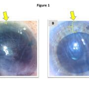
Figure 1. Despite an identical graft diameter of 8 mm in both cases, the graft is grossly oversized in this small eye with a horizontal white-to-white measurement of 11.3 mm (a) and grossly undersized in this eye with horizontal white-to-white of 13.1 mm (b). Note particularly the nasal aspect of the transplant in both instances (yellow arrow), which is positioned nearly inside the nasal angle in the first case (a), but with approximately 3 mm of separation in the second (b).

This obligation to specify in advance may result in a one-size-fits-all solution in which surgeons commonly use a standard/default graft size for (nearly) all patients. However, this default may occasionally produce undersized grafts, which inadequately treat the patient’s endothelial dysfunction (Figure 1a), or oversized grafts, which are more difficult to unfold and potentially prone to detachment (Figure 1b).

Rather than default sizes, it therefore may be desirable to tailor graft diameters for individual patients. However, this would require a method for reliably and conveniently measuring dimensions of the recipient eye before surgery. To this end, we propose a simple strategy — namely, using the preoperative horizontal corneal white-to-white (HWTW) provided by the Pentacam HR (Oculus).

The Pentacam HR employs rotating Scheimpflug imaging to obtain 50 images of potentially 138,000 corneal locations, thereby generating data for the HWTW, which may be used to calculate the graft diameter necessary to cover the recipient posterior corneal surface.

Recently, we retrospectively evaluated our last 74 consecutive DMEK operations using “patient ready” tissue. Then, we compared our subjective surgical impression of the size of the graft (well sized vs. too big vs. too small) against the preoperative Pentacam HR measurements of HWTW to generate a graft sizing nomogram, which is presented in the table.

Corneal HWTW (mm) Graft diameter (mm)
< 10.5 7
10.5 to 10.75 7.25
10.75 to 11 7.5
11 to 11.25 7.75
11.25 to 11.5 8
11.5 to 11.75 8.25
11.75 to 12 8.5
12 to 12.25 8.75
> 12.25 9

In our cohort of 74 eyes, the median HWTW was 11.65 mm, with 25% of eyes falling below 11.3 mm and 25% above 11.8 mm. For the median HWTW, a graft diameter of 8.25 mm appeared to be the optimal size (ie, could be unfolded easily and applied to the posterior cornea with a small peripheral rim of unstripped host Descemet’s membrane without overlapping). For eyes with HWTW higher or lower than the median value, the graft diameter should be adjusted, as described in the table.

Since implementing this nomogram, our incidence of mis-sized grafts has significantly decreased, resulting in easier and more enjoyable operations and fewer postoperative detachments. By incorporating more data points or additional patient anatomic parameters (for example, anterior chamber depth), it may be possible to further refine this nomogram and to provide detailed recommendations about graft sizing in difficult eyes (for example, eyes with very shallow or deep anterior chambers).

Abandoning stamp can make DMEK better

S- or F-stamping donor tissue for Descemet’s membrane endothelial keratoplasty is widely practiced to guard against DMEK’s most embarrassing potential complication: accidental upside-down graft implantation.

These orientation marks are so common, in fact, that many DMEK surgeons have never even attempted the operation without them. However, the originally described DMEK technique was stamp-free, and that is still the way the operation is performed today at the Netherlands Institute for Innovative Ocular Surgery, where DMEK was invented. So, rather than a necessary feature, tissue stamps might be more appropriately regarded as “accessories,” with definite yet unmentioned drawbacks.

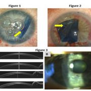
Figure 1. DMEK graft immediately after injection. Unfolding must initially proceed without use of the S-stamp. Figure 2. S-stamp only partially visible secondary to inadequate/overly tentative application. Figure 3. Anterior segment OCT sections 1 day after DMEK and corresponding slit lamp photos. In the superior cornea, the graft is well attached, and the overlying stroma is thin and clear. However, inferiorly in the location of the S-stamp, the stroma is edematous, suggesting local dysfunction of the graft/endothelium.

Admittedly, a certain emotional sense of security accompanies the use of stamped tissue. It feels like the safe thing to do. And if applied by the eye bank, it also requires no extra time, effort or expense. Finally, when not using stamps, the inevitable occurrence of an upside-down graft is so chastening and humiliating that it is enough to send any reasonable person searching for a “never again” solution.

But, in practice, the stamp is just not that useful. The DMEK graft is almost always injected as a scroll, then opened and manipulated, even before the stamp is remotely discernable (Figure 1). Consequently, the stamp has no role in the critical part of the surgery (unfolding the graft); it only offers a final check against mistakes. But even this function is often thwarted because, frequently, the stamp is only partially applied, such that half of the common “S” or “F” is missing, rendering it nearly impossible to read (Figure 2). The reason these stamps are often so poorly retained is that eye banks are typically tentative in applying them because the process (ie, stamping with a metallic instrument dipped in ink and alcohol) is unquestionably damaging, resulting in a circular swath of dead graft. These areas are additionally liable to local detachment and delayed corneal clearance (Figure 3), and the ink marks remain faintly visible for years after the surgery. As a result, the typical outcome is a poor stamp, useless for determining orientation but visible forever in the patient’s eye in a region of dead cells.

Jack S. Parker

It is also worth remembering that upside-down graft implantation is rare, occurring in fewer than 5% of operations, even without the use of tissue stamps and even during initial learning curves. So, because the potential benefit is (at most) one out of 20 cases, that means 19 out of 20 cases can only be harmed by the application of the stamp, which destroys cells every single time.

Finally, one of the most personally satisfying elements of DMEK is the beauty and elegance of the operation, which is partially spoiled by an unseemly purple tattoo. Abandoning the stamp might not make the operation easier, but it does make it better, and if DMEK surgeons did not care about that, then they would not have switched over from Descemet’s stripping automated endothelial keratoplasty in the first place.

Reference:

Dirisamer M, et al. Am J Ophthalmol. 2011;doi:10.1016/j.ajo.2011.03.031.

Ring of trypan blue aids visualization in DMEK

When starting with Descemet’s membrane endothelial keratoplasty, it is common to manually mark a ring of purple dots on the corneal surface to serve as a centering guide for the graft’s final position.

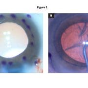
Although technically easy to apply, this array of purple dots may entail two notable drawbacks. First, inking the corneal surface is a somewhat tedious process and, while not inordinately time-consuming, nevertheless represents some period of extra manipulation. Second and more significantly, these purple dots may obscure the surgeon’s view of the most important part of the graft (its far edges), especially because ink marks are not easily applied precisely and may tend to bleed and run across the corneal surface (Figure 1a).

Figure 1. Purple dots (a) are tedious to apply and bleed together, obscuring visualization. In contrast, the blue ring (b) is subtle and clear.

Source: Jack S. Parker, MD, PhD

As a result, while the purple dots may offer some benefit, they also involve extra effort and may worsen intraoperative visualization of the most important locations. Recently, we have discovered an alternative to the purple dots that is quicker and easier to apply and also avoids hindering the surgeon’s view of the underlying area.

Intraoperatively, after trephination of the graft to size, the same trephine is used to gently create a light circular impression on the recipient corneal epithelium. The cornea is then dried, and several drops of VisionBlue (trypan blue ophthalmic solution 0.06%, DORC International) are applied. This generates a vanishingly thin blue ring on the corneal surface, exactly the same size and shape as the graft (Figure 1b), which may serve as a useful landmark, both for stripping the recipient Descemet’s membrane and for centering the donor tissue.

Compared with the purple dots, the blue ring is more precise and more subtle. It is also less laborious to apply and less likely to interfere with visualization of the graft edges. As a result, it may permit better graft centration and easier identification of abnormalities at the graft edges, resulting in a theoretically reduced risk for detachment and improved postoperative outcomes.